

That spot on your shoulder has been there a while. Is it new? Has it changed? You cannot quite remember, and that uncertainty is exactly why it’s worth a closer look. So how to know if you have skin cancer starts with knowing what to look for.
Inside this guide:
- The ABCDE rule for suspicious moles
- What each type of skin cancer looks like
- How to do a monthly self-exam
- Who faces the highest risk
- When to see a dermatologist now
Natura offers professional skin examinations to assess concerning lesions and discuss individual risk. Self-checks and professional exams can help identify changes, but neither detects every cancer.
Why Prompt Evaluation Matters
Melanoma confined to its original site generally has a better outlook than melanoma that has spread. Population survival statistics are not cure rates or individual predictions. Prognosis also depends on tumor biology, stage, overall health and response to treatment.
If a spot is new, changing, bleeding or not healing, arrange an assessment rather than waiting for the next routine check. Photos may help document a change but do not replace evaluation.
The ABCDE Rule Explained
Dermatologists built this memory tool to help patients spot melanoma early. Run any concerning mole through these five checks.
| Letter | What to look for |
|---|---|
| A: Asymmetry | One half of the mole does not match the other half |
| B: Border | Edges look irregular, notched, scalloped, or blurred |
| C: Color | Multiple colors in one spot: brown, black, tan, red, white, or blue |
| D: Diameter | Melanomas are frequently more than 6 mm across, although some can be smaller than this. |
| E: Evolving | Any change in size, shape, color, or elevation over time |
Why E matters most
Of the five, evolution carries the most weight. A mole that changes in size, shape, color, or elevation, or that starts itching, bleeding, or crusting, deserves prompt evaluation. Normal moles stay stable for years. Change is the signal.
The ugly duckling sign
There’s a second tool worth knowing. If you have many moles, look for the one that stands out from the rest. Providers call this the “ugly duckling” sign: one spot that looks different from your others, perhaps scabbed, larger, or more raised.
Your moles should mostly resemble each other. The outlier earns a closer look, even if it passes the ABCDE checks. Our guide on what a mole on skin actually is helps you recognize what normal looks like on your body.
An honest limitation
The ABCDE rule is powerful, but not perfect. Some melanomas do not meet all the criteria, and some harmless moles look suspicious. That’s precisely why dermatologist evaluation, not a home checklist, delivers the actual answer.
Warning Signs by Cancer Type
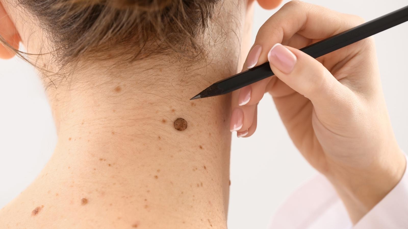
Skin cancer is not one disease. The three main types look different, so knowing each helps you spot what a mole-focused check might miss.
Basal cell carcinoma
This is the most common type of skin cancer. Basal cell carcinoma tends to grow slowly and rarely spreads, but it still needs treatment. Watch for:
- A pearly or waxy bump, sometimes skin colored or pink
- A flat, flesh-colored or brown scar-like lesion
- An open sore that bleeds, scabs, and returns
- A spot that never fully heals
These often appear on sun-exposed areas like the face, ears, and neck. Our basal cell carcinoma page covers treatment options in depth.
Squamous cell carcinoma
The second most common type, with an estimated 1.8 million diagnoses annually in the U.S. Signs include:
- A rough, scaly patch that may be reddish
- A firm, raised bump that can feel painful
- An open sore that persists or bleeds
- A rough patch on the lower lip or ear
Squamous cell carcinoma can spread if ignored, so early evaluation matters. Learn more about squamous cell carcinoma treatment.
Melanoma
The least common of the three, and the most dangerous form. Melanoma can develop from an existing mole or appear as an entirely new growth on normal skin.
Melanoma warning signs follow the ABCDE pattern above. It can also show up in unexpected places, including under fingernails, on the soles of the feet, and in areas that rarely see sun. Understanding melanoma stages clarifies why catching it early changes everything.
Signs common to all types
Regardless of type, these symptoms warrant a call:
- A spot that bleeds without an obvious injury
- A sore that does not heal within two to four weeks
- A growth that itches, stings, or hurts
- Redness or swelling spreading beyond a spot’s border
How to Do a Skin Self-Exam
Regular self-examination can help you notice changes, but it cannot diagnose or exclude skin cancer. The routine takes about ten minutes once you get the hang of it.
Your step-by-step check
- Set up. Stand in a well-lit room with a full-length mirror and a hand mirror.
- Face and scalp. Check your face, ears, lips, and neck. Part your hair in sections or use a blow dryer to inspect your scalp.
- Upper body. Examine your chest, underarms, both sides of your arms, palms, and between your fingers, including under the nails.
- Back and shoulders. Use the hand mirror against the full-length mirror, or ask a partner for help with hard-to-see areas.
- Lower body. Check your abdomen, buttocks, both sides of your legs, soles of your feet, and between your toes.
- Document. Photograph a concerning spot for your clinician and arrange an assessment; do not wait a month to seek advice about a new or changing lesion.
Do not skip hidden spots
Skin cancer can develop in areas that never see sunlight. That means checking your scalp, the soles of your feet, under your nails, and skin folds. These spots get overlooked, and cancers there are often found late.
A note on skin tone. Skin cancer affects every complexion. On black skin and darker tones, melanoma more often appears on palms, soles, and under nails, and it’s frequently diagnosed later. Our guide on whether black people can get skin cancer addresses this directly.
Who Faces Higher Risk
Anyone can develop skin cancer, but certain factors raise your odds considerably. Knowing where you stand helps you decide how often to get screened.
| Risk factor | Why it increases risk |
|---|---|
| Heavy sun exposure | UV radiation damages skin cell DNA over time |
| Tanning bed use | Concentrated ultraviolet light accelerates damage |
| Fair skin, light hair | Less melanin means less natural UV protection |
| Family history | Genetics influence susceptibility |
| Many moles | More moles, more opportunity for change |
| Weakened immune system | Reduced ability to fight abnormal cells |
| Prior skin cancer | History raises the odds of recurrence |
If several apply to you, ask your dermatologist about a regular screening schedule. High-risk patients benefit from professional exams even when nothing looks obviously wrong.
Prevention still works
Reducing risk is straightforward, and it starts with sun protection:
- Wear broad-spectrum sunscreen SPF 30 or higher daily
- Reapply every two hours when outdoors, more often if swimming
- Seek shade between 10 a.m. and 2 p.m. when UV rays peak
- Wear protective clothing and a wide-brimmed hat
- Skip tanning beds entirely
Our sunscreen collection makes daily protection easier, and our guide on how to avoid skin cancer goes deeper on prevention strategy.
When to See a Dermatologist
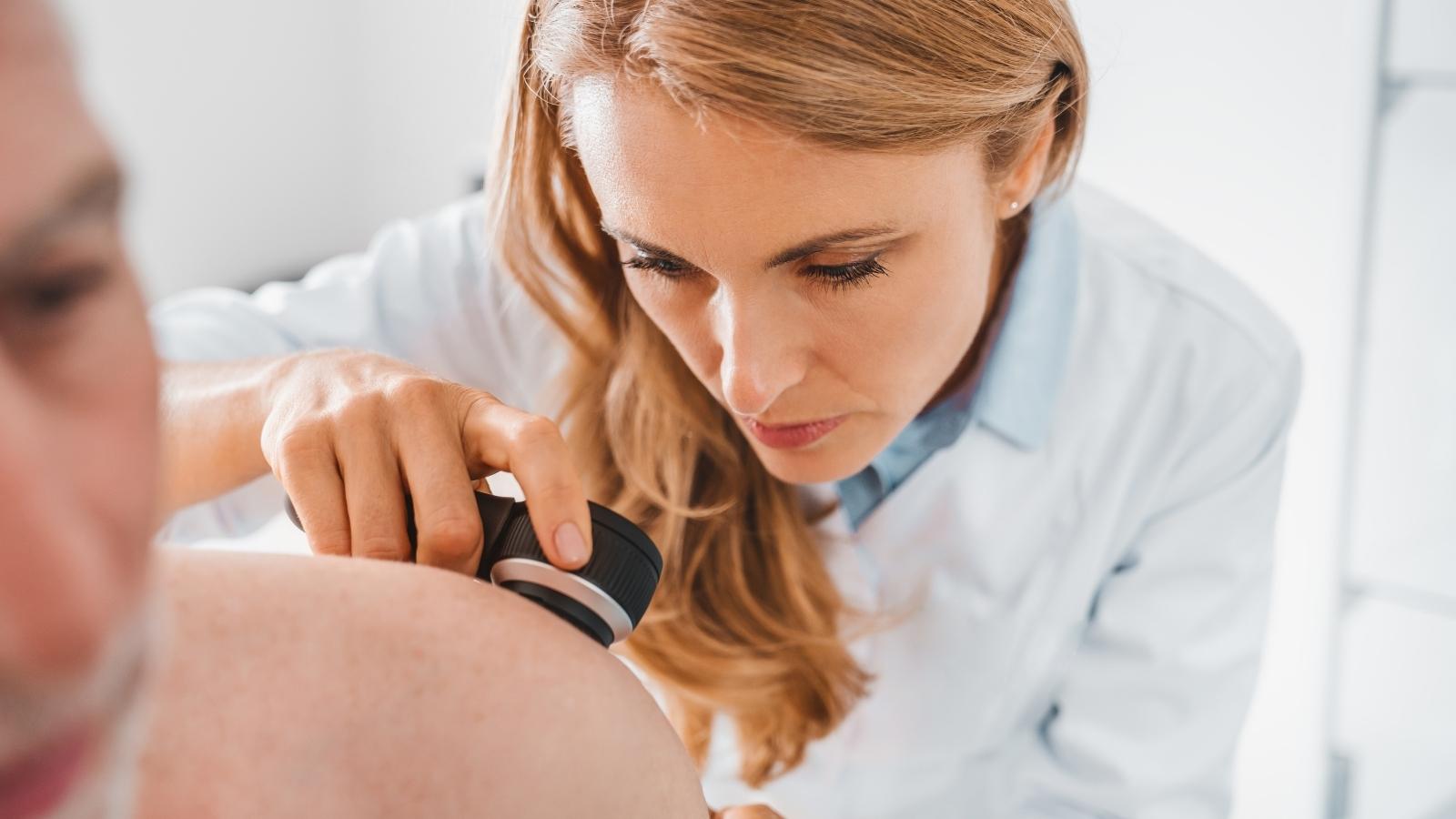
Here’s the simplest guidance in this entire article: when in doubt, get it checked. A false alarm costs you an appointment. Delay can cost far more.
Book an evaluation right away if you notice:
- A mole that meets any ABCDE criteria
- A spot that looks different from your other moles
- A sore that has not healed in two weeks or more
- Any growth that bleeds, itches, or changes
- A new mole appearing after age 30
What happens at your appointment
Knowing the process removes the anxiety. Your dermatologist examines your skin closely, often with a dermatoscope for magnification. If a spot looks suspicious, they perform a skin biopsy, removing a small sample for lab analysis.
That biopsy is what confirms or rules out cancer. Diagnosis requires a physical exam with a dermatologist plus a biopsy, and only then can treatment be recommended. If treatment is needed, options range from simple excision to Mohs surgery, which removes cancer precisely while preserving healthy tissue.
Do not let fear delay you. Early evaluation significantly improves outcomes, and most suspicious spots turn out to be nothing. Our guide on when to see a dermatologist covers other signs worth acting on.
What Skin Cancer Is Not
Not every new spot spells trouble. Plenty of harmless growths mimic the warning signs, and knowing the difference saves you unnecessary worry. It also keeps you from dismissing something that genuinely needs a doctor.
Common benign lookalikes
Most skin growths people find are not cancer at all. These show up constantly:
| Growth | What it looks like |
|---|---|
| Skin tag | Soft, flesh-colored flap, often in skin folds |
| Seborrheic keratosis | Waxy, stuck-on brown patch, harmless despite the look |
| Common moles | Round, even color, stable for years |
| Cherry angioma | Small bright red dot from blood vessels |
| Dry skin patches | Flaky, itchy skin that improves with moisturizer |
Dry skin deserves a mention here because it fools people often. A rough, itchy patch that responds to moisturizer within a week or two is usually just dryness. One that persists, bleeds, or spreads is not.
Common moles stay consistent. They hold one color, keep a smooth edge, and look like your other existing moles. A suspicious mole breaks that pattern with an irregular border, mixed colors, or steady change.
If you want a growth removed for comfort or appearance rather than concern, our skin tag removal and mole removal services handle both, with evaluation built into the visit.
How skin cancer actually develops
Understanding the mechanism explains why early treatment works so well. Skin cancers begin when UV damage causes skin cells to mutate. Those cancer cells then multiply out of control instead of dying off normally.
Early skin cancer stays contained in the upper layers, which is why removal at that stage delivers such successful treatment rates. Left alone, some types grow deeper. In rare cases, melanoma and advanced squamous cell carcinoma reach the lymph nodes and spread further, which is where outcomes change sharply.
Basal cell carcinoma rarely travels, while other skin cancers vary depending on type and depth. That difference is exactly why identifying the type matters, and why a biopsy guides treatment.
Knowing your personal risk
Not everyone faces the same odds of developing skin cancer. Your risk of developing skin problems rises with fair skin types, a family history of skin cancer, or a personal history of skin cancer. Any of these puts you in the increased risk group.
That does not mean waiting for symptoms. High-risk patients benefit from routine professional exams, since a doctor spots changes on the scalp and back that you simply cannot see. To prevent skin cancer in the first place, daily sun protection remains the most reliable tool you have.
Get a Concerning Spot Checked
Being familiar with your usual skin and having new or changing spots evaluated promptly can support earlier recognition of possible skin cancer. A professional examination cannot guarantee that every cancer will be detected, and a photograph or checklist cannot rule it out.
Ask your dermatologist about a follow-up schedule based on your history and risk. For a changing, bleeding or non-healing spot, contact Natura for an evaluation rather than waiting for a routine screening appointment.
Frequently Asked Questions
What are possible early signs of skin cancer?
A new or changing lesion, a pearly bump, a persistent scaly patch or a sore that does not heal can warrant assessment. Skin cancer may also have less typical appearances.
Is skin cancer always curable?
No. Treatment can be successful for many skin cancers, particularly when the disease is still localized, but the outlook varies according to cancer type, stage, and individual circumstances.
What does stage I melanoma look like?
Appearance cannot establish stage. Diagnosis requires tissue examination; staging uses pathology and, when indicated, lymph-node assessment or other tests.
How is skin cancer diagnosed?
A clinician examines the skin and may take a biopsy of a suspicious lesion. Home checks alone cannot confirm or exclude cancer.


