

A melanoma diagnosis hits different when you understand the stages. The difference between Stage I and Stage III isn’t just medical jargon—it’s the difference between a 99% and 63% five-year survival rate. Yet most people don’t fully grasp what their melanoma stage means until treatment decisions are already being made.
We’re breaking down exactly what happens at each stage and why these classifications matter for your treatment path.
What we’ll cover:
- The staging system and how it works
- What each stage means for spread and survival
- Key factors that determine your stage
- Treatment options by stage
- Signs of progression to watch for
At Natura Dermatology, we catch most melanomas at Stage 0 or I through our comprehensive skin checks and imaging technology. Dr. Richardson’s expertise in Mohs surgery means that even concerning cases can be completely removed while preserving healthy tissue.
The Staging System and How It Works
Understanding melanoma skin cancer stages starts with knowing how doctors classify this disease. The system isn’t arbitrary—it’s based on decades of data about how melanoma cells behave and spread.
The TNM Foundation
The National Comprehensive Cancer Network uses the AJCC Cancer Staging Manual’s TNM system:
- T (Tumor): Thickness and characteristics of primary melanoma
- N (Nodes): Lymph node involvement
- M (Metastasis): Spread to distant parts of the body
These three factors combine into stage grouping from 0 to IV, with substages providing more detail.
Key Measurements
Doctors evaluate several critical factors when determining your stage:
| Factor | What It Measures | Why It Matters |
|---|---|---|
| Breslow Thickness | Tumor thickness in millimeters | Best predictor of outcome |
| Ulceration | Broken skin over the tumor | Indicates aggressive behavior |
| Mitotic Rate | Cell division speed | Shows growth rate |
| Node Status | Cancer in the lymph nodes | Determines regional spread |
The pathological stage (determined after surgery) often differs from the clinical stage (before surgery), which is why biopsies are critical.
What Each Stage Means for Spread and Survival
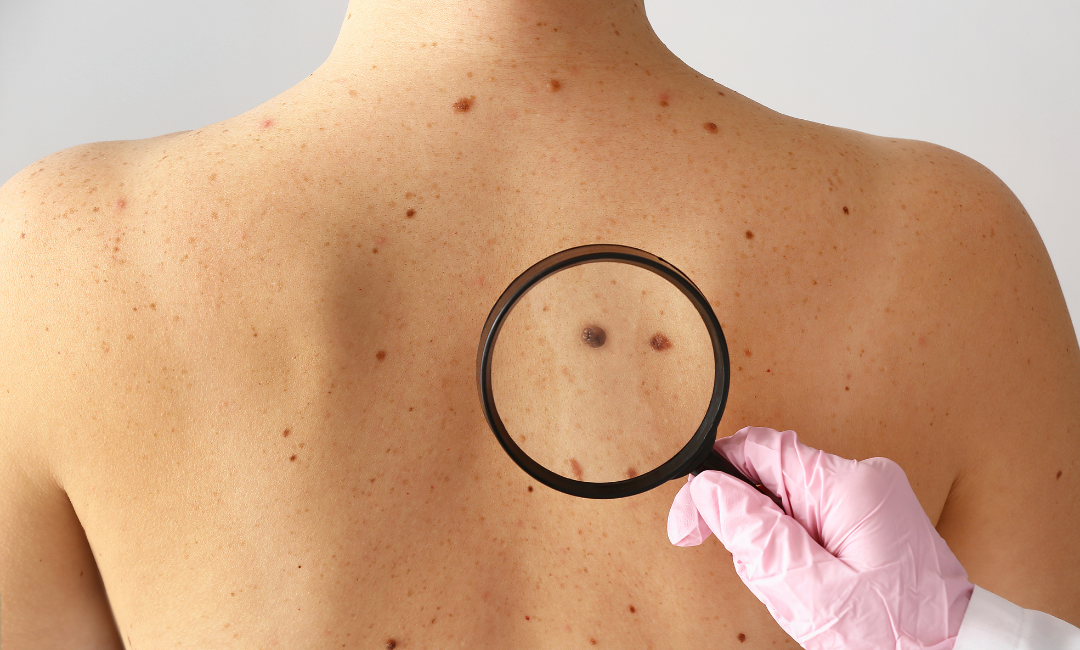
Each melanoma stage tells a different story about where cancer cells have traveled and what that means for your prognosis. Understanding these distinctions helps you grasp why treatment recommendations vary so dramatically between stages.
Stage 0 Melanoma (In Situ)
Cancer cells exist only in the epidermis—the skin’s top layer. This stage, called melanoma in situ, hasn’t invaded deeper tissue.
- Spread: None
- 5-year survival: Nearly 100%
- Treatment: Wide excision only
Stage I Melanoma
The primary tumor is thin (under 2mm) with no lymph node involvement. This early-stage diagnosis offers excellent outcomes with proper treatment.
- Stage IA: Under 0.8mm, no ulceration
- Stage IB: 0.8-1mm with ulceration, or 1-2mm without
- 5-year survival: 92-99%
- Treatment: Wide excision, possible sentinel node biopsy
Stage II
Thicker melanoma tumors characterize this stage, but there’s still no spread to nearby lymph nodes yet. The tumor thickness determines substaging.
- Stage IIA: 1-2mm with ulceration, or 2-4mm without
- Stage IIB: 2-4mm with ulceration, or over 4mm without
- Stage IIC: Over 4mm with ulceration
- 5-year survival: 53-82%
- Treatment: Wide excision, sentinel node biopsy, possible immunotherapy
Stage III Melanoma
Cancer has spread to nearby lymph nodes or nearby skin (satellite tumors or in transit tumors). This regional spread marks a critical turning point in the treatment approach.
- Substages IIIA-IIID based on the number of tumor-involved nodes
- Includes: Regional metastases, microsatellite metastases
- 5-year survival: 40-78%
- Treatment: Surgery, immunotherapy, targeted therapy, and a possible clinical trial
Stage IV Melanoma
Metastatic melanoma has reached distant lymph nodes, distant skin, or organs. Modern treatments have dramatically improved outcomes at this stage.
- Common sites: Lungs, liver, brain, bones, central nervous system
- Distant metastases classified by location
- 5-year survival: 15-30% (improving with new treatments)
- Treatment: Immunotherapy, targeted therapy, radiation, clinical trials
Key Factors That Determine Your Stage
Multiple characteristics of your melanoma work together to determine staging. Each factor provides crucial information about how aggressive the cancer might be.
Primary Tumor Characteristics
Breslow Thickness remains the most important factor. Each millimeter of depth dramatically changes prognosis:
- Under 1mm: Excellent outlook
- 1-2mm: Good with proper treatment
- 2-4mm: Moderate risk requiring aggressive treatment
- Over 4mm: High risk of spread
Ulceration indicates the tumor has broken through the skin surface. Ulcerated melanomas behave more aggressively, automatically bumping you to a higher substage.
Lymph Node Assessment
The extent of lymph node involvement significantly impacts staging and treatment decisions. Doctors evaluate:
- Sentinel nodes: First nodes cancer would reach
- Number involved: More nodes = higher stage
- Microscopic vs. macroscopic: Size of cancer deposits matters
The difference between microscopic and visible node involvement can mean moving from Stage IIIA to IIIC—a significant jump in treatment intensity.
Special Considerations
Several unique features affect staging beyond basic measurements.
- Mitotic rate (how fast cells divide) affects staging for thin melanomas.
- Satellite lesions within 2cm of the primary tumor indicate local spread.
- In-transit metastases between the primary site and nodes suggest aggressive disease.
Invasive melanoma that shows these features requires immediate, comprehensive treatment regardless of thickness.
Treatment Options by Stage

Treatment strategies shift dramatically based on your melanoma stage, with early stages requiring less intensive intervention than advanced disease.
Early Stage (0-IIA)
Treatment focuses on complete removal with clear margins:
- Wide local excision: Removes tumor plus safety margin
- Margin width: 0.5-2cm, depending on thickness
- Sentinel node biopsy: For tumors over 0.8mm
- Follow-up: Regular skin checks every 3-6 months
Intermediate Stage (IIB-IIIC)
A multi-modal approach becomes necessary when melanoma shows aggressive features or regional spread:
- Surgery: Remove primary tumor and affected nodes
- Adjuvant immunotherapy: Reduces recurrence risk by 40-50%
- Options: Pembrolizumab, nivolumab, or targeted therapy
- Monitoring: PET/CT scans every 3-4 months
Advanced Stage (IIID-IV)
Systemic therapy takes priority when cancer has spread beyond regional control:
- First-line immunotherapy: Combination checkpoint inhibitors
- Targeted therapy: For BRAF-mutant melanomas
- Radiation: For brain metastases or symptom control
- Clinical trials: Access to cutting-edge treatments
Response Rates by Treatment
Modern treatments have revolutionized outcomes, especially for advanced stages:
| Treatment Type | Response Rate | Duration |
|---|---|---|
| Single immunotherapy | 40-45% | Often years |
| Combo immunotherapy | 55-60% | Potentially permanent |
| BRAF/MEK inhibitors | 70% | 11-15 months median |
| Surgery (early stage) | 90-95% cure | Permanent if clear |
The key: catching cutaneous melanoma early means simpler treatment with better outcomes.
Navigate Melanoma Stages with Natura Dermatology
Understanding melanoma skin cancer stages empowers you to make informed treatment decisions. Whether you’re facing stage I melanoma or metastatic melanoma, knowing what each stage means transforms fear into action.
Key takeaways:
- Staging combines tumor depth, node involvement, and spread
- Early stages (0-I) have 90%+ survival rates
- Stage III melanoma varies widely in outcomes
- Stage IV treatment has improved dramatically
- Breslow thickness drives initial staging
- Regular monitoring catches progression early
Natura Dermatology specializes in early detection and comprehensive melanoma care. Our team uses advanced imaging to identify concerning lesions before they progress, and Dr. Richardson’s surgical expertise ensures complete removal when skin cancer is found.
FAQs
How long does it take melanoma to go from stage 1 to stage 4?
Progression varies dramatically—some melanomas remain stable for years while others advance within months. Aggressive subtypes like nodular melanoma can reach stage IV within 6-12 months. Most cutaneous melanomas take 2-5 years to progress through stages of melanoma if left untreated.
At what stage is melanoma not curable?
No stage is automatically incurable anymore. Even stage IV melanoma sees long-term remission in 20-30% of patients with modern immunotherapy. However, cure rates drop significantly after lymph node involvement (stage III). Early stages offer 90%+ cure rates with surgery alone.
What does a stage 4 melanoma look like?
Stage IV means the cancer has spread beyond regional lymph nodes to distant organs. The original skin lesion might be gone (removed earlier) while metastases cause symptoms elsewhere—breathing issues (lungs), confusion (brain), or pain (bones). Distant skin metastases appear as multiple dark nodules.
What does a stage 2 melanoma look like?
Stage II presents as a deeper lesion (over 1mm thick) often with irregular borders, multiple colors, and a diameter over 6mm. It may appear raised, dome-shaped, or ulcerated. The primary tumor looks concerning but shows no spread to nearby lymph nodes on examination or imaging.
What are the five stages of melanoma?
The five main stages are: Stage 0 (in situ—top layer only), Stage I (thin, localized), Stage II (thicker, still localized), Stage III (spread to nearby lymph nodes or skin), and Stage IV (distant metastases). Each has substages (IA, IB, IIA, etc.) based on specific features.
What is the life expectancy of someone with stage melanoma?
Life expectancy depends entirely on stage at diagnosis. Stage 0-I patients typically have normal life expectancy. Stage II shows 53-82% five-year survival. Stage III varies from 40-78%. Even stage IV patients now see 30% five-year survival with new treatments—and this keeps improving yearly.


