

Your face takes more sun than any other part of your body. Decades of it, whether you tanned deliberately or just drove to work. That’s why the face ranks among the most common sites for skin cancer, and why knowing the types of skin cancer on the face matters so much.
Inside this guide:
- The three main facial skin cancers
- Rare types worth knowing
- Where each one typically appears
- Treatment options by type
- When to book an appointment
Natura evaluates suspicious skin changes and helps patients understand their individual risks and treatment options. Prompt assessment matters, but no examination guarantees detection of every cancer.
Why the Face Is So Vulnerable
Your face rarely gets a day off from ultraviolet light. Skin cancers frequently develop on sun-exposed areas, and the face, nose, ears, and lips top that list.
Basal cell carcinomas most commonly appear on sun-exposed skin, especially the face, while squamous cell carcinomas typically show up on the face, ears, lips, neck, and backs of the hands.
Here’s what makes facial skin cancer distinct:
- Cumulative exposure builds over decades of ordinary daily life
- Thin skin on eyelids, nose, and ears offers less protection
- Cosmetic stakes are higher, since treatment can leave visible scarring
- Early detection matters doubly, reducing both spread and disfigurement
The upside: facial spots are easy to see. You look at your face daily, which gives you a real advantage over cancers hiding on your back or scalp.
Pro tip: Photograph a concerning spot to document its appearance and arrange an assessment. If a spot is new, evolving, bleeding, or failing to heal, have it evaluated promptly rather than waiting several weeks.
Basal Cell Carcinoma on the Face
This is the one you’re most likely to encounter. Basal cell carcinoma is the most common type of skin cancer, and the face is its favorite address.
What it looks like
Basal cell carcinomas often appear as a flesh-colored round growth, a pearl-like bump, or a pinkish patch of skin. Watch for:
- A pearly or waxy bump, sometimes with visible tiny blood vessels
- A flat, scar-like lesion that is flesh-colored or brown
- An open sore that bleeds, crusts over, and returns
- A pink growth with a slightly raised, rolled border
Where it appears
Common facial locations include the nose, forehead, cheeks, eyelids, and ears. The nose is a particularly frequent site.
How serious is it
Basal cell cancers grow slowly and rarely spread to other parts of the body. That’s the reassuring part. But left untreated, they can grow into nearby areas and invade bone or other tissues beneath the skin.
There’s also a recurrence pattern worth knowing. Within five years of a basal cell diagnosis, a substantial share of patients develop another skin cancer. One diagnosis means ongoing monitoring, not a one-and-done fix. Our basal cell carcinoma page covers treatment in detail.
Squamous Cell Carcinoma on the Face
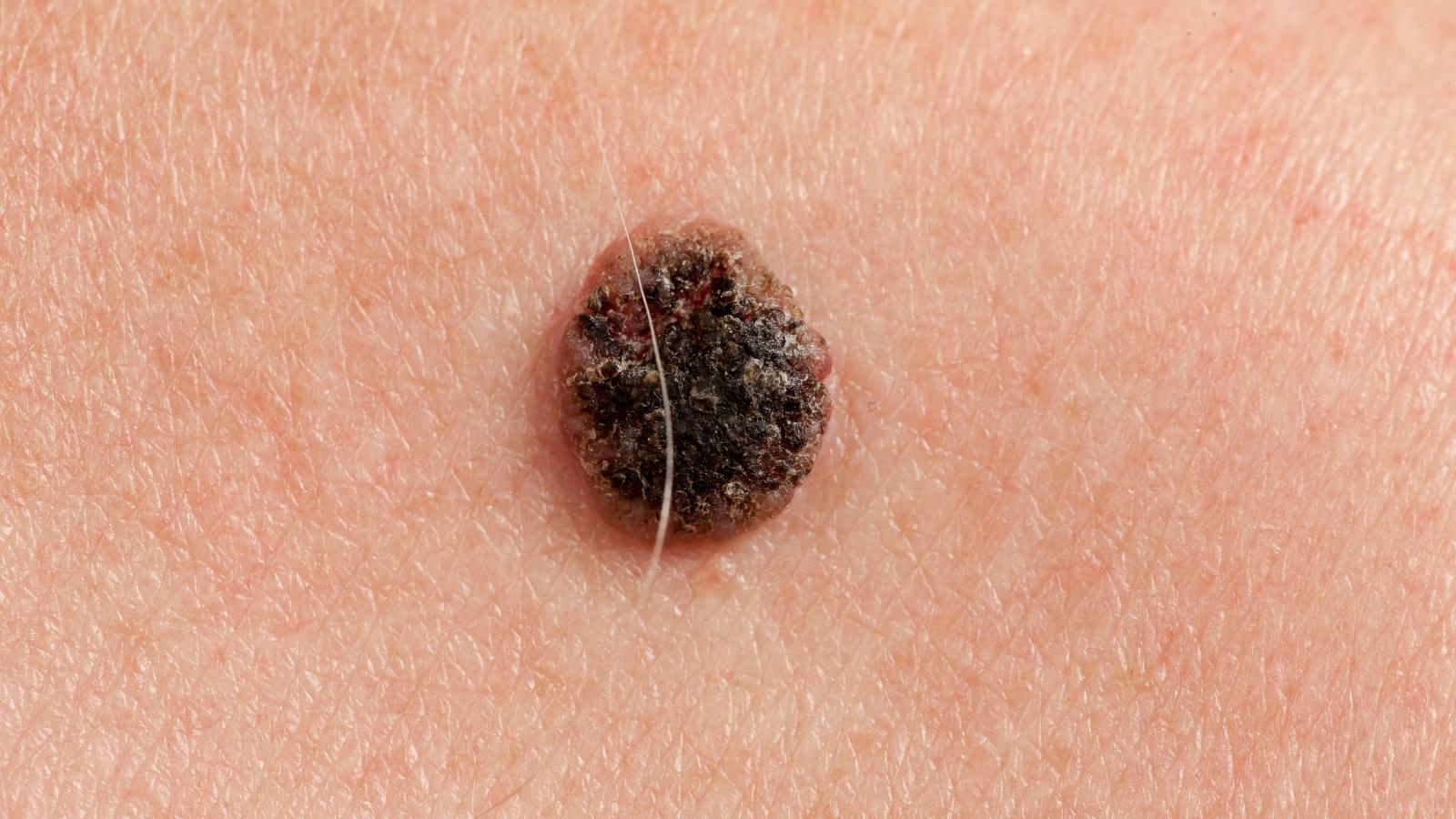
The second most common type, and more assertive than its basal cell cousin. About 2 out of 10 skin cancers are squamous cell carcinomas, which start in the flat cells of the outer epidermis.
What it looks like
Squamous cell skin cancer tends to announce itself with texture:
- A rough, scaly patch that may crust or bleed
- A firm red nodule or raised bump
- A wart-like growth with a crusted surface
- An open sore that will not heal
- A rough patch on the lower lip or the rim of an ear
Where it appears
Squamous cell cancers commonly appear on the face, ears, neck, lips, and backs of the hands. Lips and ears deserve special attention, since cancers there carry a higher risk of spreading.
How serious is it
Squamous cell carcinomas are generally more aggressive than basal cell cancers. They can travel to nearby lymph nodes, particularly when they arise on the lip or ear, or in people with a weakened immune system.
Some squamous cell carcinomas develop from actinic keratoses, which are precancerous lesions. Treatment can reduce risk at treated sites, but it cannot prevent every future skin cancer. Continued sun protection and follow-up remain important.
Melanoma on the Face
The least common of the three main types, and the one that demands the fastest action. Malignant melanoma begins in melanocytes, the cells that produce your skin’s pigment.
What it looks like
Melanoma can grow from an existing mole or appear as an entirely new dark spot. Use the ABCDE rule:
| Letter | Warning sign |
|---|---|
| A: Asymmetry | One half does not match the other half |
| B: Border | Edges look irregular, notched, or blurred |
| C: Color | Multiple shades of brown, black, tan, red, or white |
| D: Diameter | Melanoma is frequently more than 6 mm across, although some lesions may be smaller than that. |
| E: Evolving | Changing in size, shape, color, or elevation |
A facial-specific pattern
Facial melanomas often behave differently from those elsewhere. Melanomas on the head and face are commonly the lentigo maligna type, associated with frequent moderate chronic UV exposure, unlike trunk melanomas linked to intense intermittent burning.
Lentigo maligna typically appears as a flat, freckle-like brown patch that slowly expands, often on the cheek or temple. Because it looks like an age spot, people frequently dismiss it. Our guide on age spots versus skin cancer helps you tell them apart.
How serious is it
Melanoma is the least frequent of the three common skin cancers, but it metastasizes readily and can cause death once it spreads. Early detection changes the outlook dramatically, which is why understanding melanoma stages matters.
Less Common Facial Skin Cancers and Precancerous Lesions
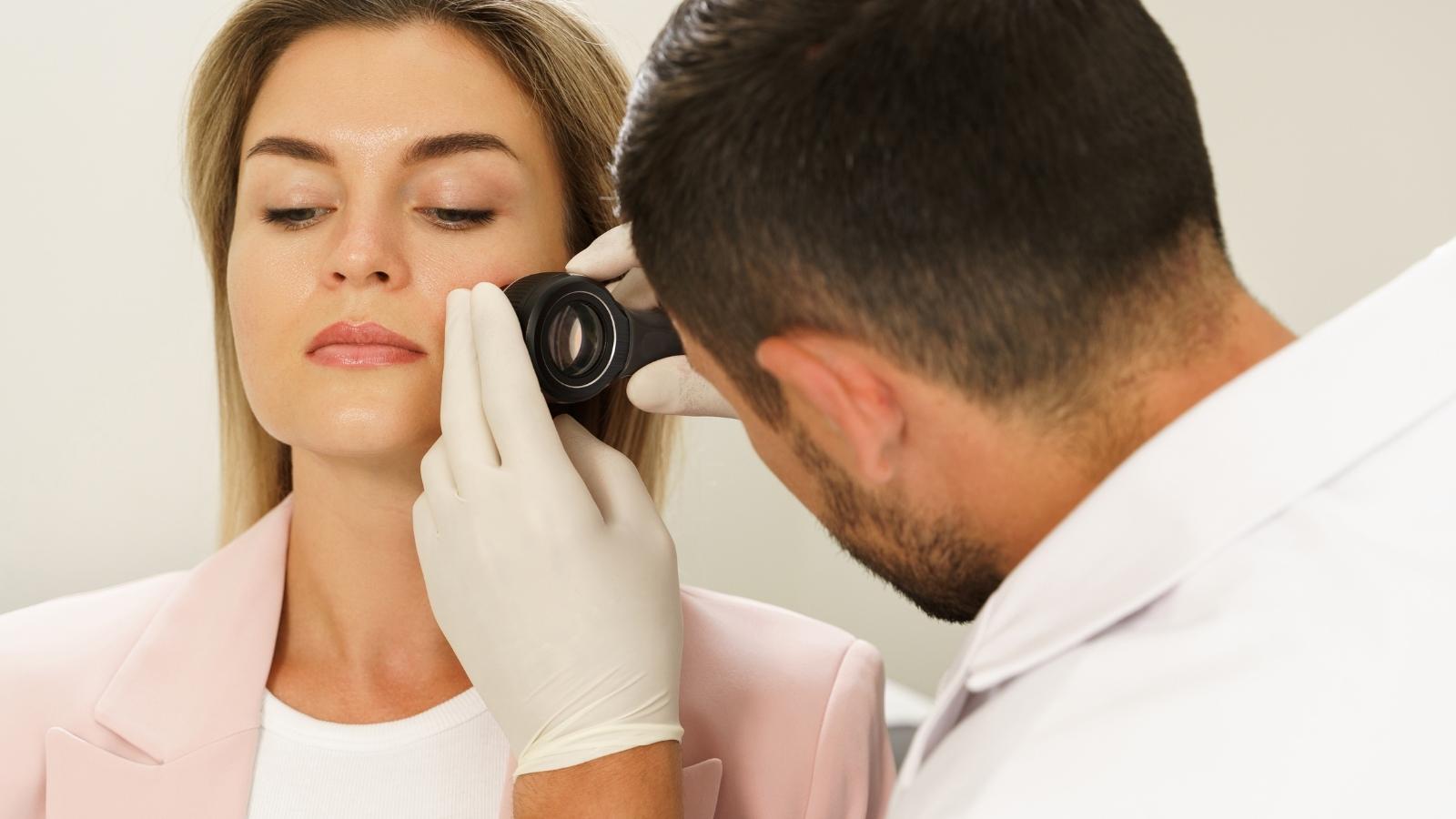
Less common cancers include Merkel cell carcinoma, sebaceous carcinoma and Kaposi sarcoma. Their appearances vary, and a persistent or rapidly changing lesion requires assessment.
Actinic keratosis is a precancerous lesion, not a type of skin cancer. It may appear as a rough or scaly patch on sun-exposed skin, and some lesions progress to squamous cell carcinoma. A dermatologist can advise which treatment and follow-up are appropriate.
Who Faces Higher Risk
Anyone can develop facial skin cancer, but certain factors raise your odds. Knowing yours helps you decide how often to get screened.
- Fair skin, light-colored eyes, red or blonde hair offer less natural UV protection
- Heavy sun exposure or a history of blistering sunburns
- Tanning bed use, which concentrates ultraviolet light
- Family history of skin cancer
- Many moles or unusual existing moles
- Weakened immune system, including immunosuppressant medications
- Prior skin cancer, which raises recurrence odds sharply
Skin cancer in darker skin tones
A dangerous myth persists that darker skin does not get skin cancer. It does. People with skin of color develop basal cell carcinoma too, though it may look different, often appearing brown or glossy black rather than pearly pink.
Melanoma in darker-skinned people more often appears on palms, soles, and under nails, and it’s frequently diagnosed later, when outcomes are worse. Our guide on whether black people can get skin cancer covers this in depth.
Treatment Options by Type
Treatment depends on the type, size, depth, and location of the cancer. On the face, preserving healthy tissue matters as much as removing the cancer.
Surgical approaches
Surgery remains the foundation of skin cancer treatment. Options include:
- Mohs surgery, removing cancer layer by layer while checking margins under a microscope
- Standard excision, cutting out the growth plus a margin of surrounding skin
- Curettage and electrodesiccation, scraping and burning small superficial cancers
- Cryotherapy, freezing off precancerous spots and some early lesions
Why Mohs matters on the face
For facial cancers, Mohs surgery is often the treatment of choice, especially for basal cell carcinoma. It removes cancer cells precisely while sparing the maximum amount of healthy tissue, which protects both function and appearance in cosmetically sensitive areas like the nose, eyelids, and lips.
Other treatments
For advanced or specific cases, additional options come into play:
- Radiation therapy for aggressive squamous cell carcinoma or patients who cannot have surgery
- Immunotherapy for advanced melanoma and some other skin cancers
- Topical chemotherapy creams for superficial lesions and actinic keratosis
- Targeted therapy for advanced melanoma with specific genetic markers
Earlier diagnosis often allows more localized treatment, but the procedure, scarring and outcome depend on the cancer’s type, site, extent and other risk features. Our skin cancer services walk through what each approach involves.
How Facial Skin Cancer Develops and Is Assessed
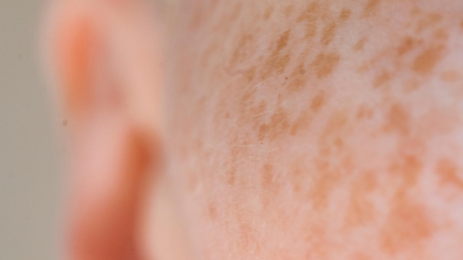
Sun-related ultraviolet damage contributes substantially to many common skin cancers, but other factors can also play a role. Each cancer type differs in how it develops, grows, and which risks are associated with it.
Skin cancers are not all classified with one universal staging system, because the criteria vary by cancer type. Melanoma staging uses features such as thickness, ulceration and spread. Squamous cell carcinoma has its own risk and staging criteria; most basal cell carcinomas are managed by local risk features rather than a routine stage 0–IV sequence. Your healthcare provider can clarify which staging or risk-assessment system applies to your specific diagnosis.
Use sun protection, avoid tanning beds and seek assessment for a new, changing, bleeding or non-healing lesion. Sunscreen reduces UV exposure but does not eliminate skin-cancer risk.
Get Your Face Checked at Natura
The types of skin cancer on the face come down to three common culprits plus a few rare ones, and every single one responds better when caught early. Your face is visible daily, which makes you your own best early warning system.
Keep these takeaways handy:
- Basal cell looks pearly and grows slowly, rarely spreading
- Squamous cell looks rough and scaly, and can spread from lips or ears
- Melanoma follows the ABCDE pattern and demands urgent evaluation
- Facial melanoma often mimics an age spot that slowly expands
- Mohs surgery preserves healthy tissue in cosmetically sensitive areas
Noticed a spot that will not heal? Book a skin cancer screening at Natura Dermatology in Fort Lauderdale, where our board-certified team performs thorough exams with advanced imaging and offers Mohs surgery on site. One appointment brings answers.
Frequently Asked Questions
Which is more serious, basal cell or squamous cell skin cancer?
Squamous cell carcinoma is typically more likely to spread beyond the original site than basal cell carcinoma. Both can cause serious local damage, and the risk of an individual tumor depends on its features and location.
What is the most common type of skin cancer in the face?
Basal cell carcinoma is the most common facial skin cancer. It typically appears as a pearly bump or pinkish patch on the nose, cheeks, or forehead.
What is the 2 week rule for skin cancer?
Any sore, scab, or spot that has not healed within two weeks should be evaluated by a dermatologist. Non-healing lesions are a key warning sign.
How to tell if it’s skin cancer face?
Watch for pearly bumps, rough scaly patches, sores that will not heal, or moles that change. Only a dermatologist can confirm through examination and biopsy.


