

Most people have between 10 and 40 moles by adulthood. The vast majority are completely harmless. But here’s what most people don’t know: roughly 1 in 100 melanoma cases develop from an existing mole. That’s a small number, but the stakes are high enough to pay attention.
This guide breaks down everything you need to know about skin moles so you can tell the difference between “totally normal” and “time to call a dermatologist.”
Here’s what we’ll cover:
- What a mole actually is and why it forms
- The different types of moles (common, atypical, congenital)
- Warning signs that a mole could be dangerous
- The ABCDE rule for self-checks
- When to see a dermatologist
- How moles are removed and what to expect
At Natura Dermatology & Cosmetics, our providers perform thorough full-body skin exams to catch potential issues early. If you’ve got a mole that’s changed or one you’re unsure about, our Fort Lauderdale team is just a call away.
What a Mole Actually Is and Why It Forms
A mole is a small growth on the skin that forms when cells called melanocytes cluster together instead of spreading evenly across your skin’s surface. Melanocytes are the cells responsible for producing melanin, the pigment that gives your skin its natural color. When they group up in one spot, you get a visible mark that can be flat or raised, and typically brown or black in shade.
Most moles appear during childhood and adolescence. Most people develop between 10 and 40 skin moles by adulthood. After age 40, new moles become less common, and existing moles tend to slowly fade or change in appearance over time. That’s normal.
Why Do Moles Form?
Two main factors drive mole development:
- Genetics. If your parents had many moles, you’re likely to develop more yourself. Family history plays a significant role in both the number and type of moles you’ll have.
- Sun exposure. UV rays stimulate melanocytes to produce more pigment as a protective response. Over time, this can trigger melanocytes to cluster, forming new moles. People with fair skin and those who spend long periods in the sun or use tanning beds tend to develop more moles.
It’s worth noting that moles can show up anywhere on the body, including areas rarely exposed to sunlight, like the scalp, between toes, or even on the lower legs. This suggests that hormones and other biological processes also play a role beyond UV rays alone.
Types of Moles You Should Know
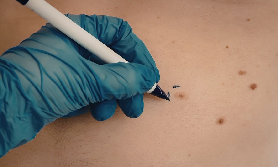
Not all skin moles look or behave the same way. Understanding the different types helps you recognize what’s normal and what deserves a closer look.
Common Moles (Acquired Nevi)
These are the moles that appear after birth, typically during childhood. A common mole is usually a small growth, less than 5 millimeters wide (about the size of a pencil eraser). It has a round or oval shape with a distinct edge and an even color, usually pink, tan, or dark brown, depending on your skin tone.
Key traits of normal moles:
- Symmetrical shape
- Smooth, well-defined borders
- Uniform color throughout
- Smaller than 6mm in diameter
- Stable over time
Most common moles are completely harmless moles that will never cause problems.
Atypical Moles (Dysplastic Nevi)
Atypical moles are larger than common moles and irregularly shaped. They often display uneven color, with a dark brown center and lighter, uneven edges that may blend into the surrounding skin. Dysplastic nevi can appear anywhere, including areas not exposed to the sun.
Here’s why they matter: the National Cancer Institute notes that people with five or more atypical moles have roughly 10 times the risk of developing melanoma compared to someone with none.
Having atypical moles doesn’t mean you have cancer. But it does mean you should be monitoring them closely.
Congenital Moles (Congenital Nevi)
Congenital nevi are moles present at birth, occurring in about 1 in 100 babies. They can range from small marks to large skin lesions covering significant areas. While most are benign, a mole larger than 8 millimeters carries a greater risk of becoming cancerous over time and should be checked regularly by a dermatologist.
Blue Moles
Blue moles get their unusual color because the melanocytes sit deeper within the skin. They appear blue or bluish-gray rather than the typical brown or black. Blue moles are generally benign, but their unusual appearance can sometimes be mistaken for something more serious, so they warrant a professional eye.
| Mole Type | Appearance | Size | Risk Level |
|---|---|---|---|
| Common | Round, even color, distinct edge | Under 5mm | Low |
| Atypical | Irregular shape, uneven color, uneven edges | Over 5mm | Moderate |
| Congenital | Present at birth, variable | Varies widely | Low to moderate |
| Blue | Bluish-gray, deeper pigment | Usually small | Low |
Warning Signs That a Mole Could Be Dangerous
Most moles are harmless. They’ll sit on your skin for decades without causing a single problem. But melanoma, the most aggressive type of skin cancer, also develops in melanocytes. That’s why knowing the difference between a normal mole and a potentially cancerous one matters.
Red Flags to Watch For
Pay attention if you notice any of these skin changes in an existing mole or a new growth:
- A mole that has changed size, shape, or color over a few weeks or months
- Bleeding or oozing without any injury
- A mole that becomes painful, itchy, or tender
- Multiple colors within a single mole (brown, black, red, white, or blue)
- Borders that become blurred, jagged, or irregularly shaped
- A new mole appearing after age 40, especially one that looks different from your others
The “Ugly Duckling” Rule
This is one of the simplest ways to spot trouble. Look at all the moles on your body. Most of them should look similar to each other. If one mole stands out as clearly different, that’s your “ugly duckling,” and it deserves a professional medical review.
A black mole among brown ones. A large, raised mole when all your others are flat. Anything that doesn’t match the pattern. Trust your instinct. If something looks off, get it checked.
The ABCDE Rule for Self-Checks

Dermatologists recommend the ABCDE method as a simple framework for evaluating skin moles at home. It won’t replace a professional exam, but it gives you a reliable system for knowing what to look for.
| Letter | What It Stands For | What to Look For |
|---|---|---|
| A | Asymmetry | One-half of the mole doesn’t match the other half |
| B | Border | Edges are irregular, blurred, or jagged |
| C | Color | Uneven color or multiple colors (brown, black, red, white, blue) |
| D | Diameter | Wider than a pencil eraser (about 6mm) |
| E | Evolving | Size, shape, color, or feel have changed over time |
How to Do a Monthly Self-Check
Set aside 10 minutes once a month. Stand in front of a full-length mirror in good lighting and do a head-to-toe check.
- Start with your face, including your ears and hairline
- Check your scalp using a handheld mirror (or ask someone to help)
- Examine your neck, chest, and torso, front and back
- Look at both arms, including underarms and palms
- Check your lower legs, ankles, and the soles of your feet
- Don’t skip between your toes or the backs of your knees
Pro tip:Take photos of any moles you’re unsure about, with a ruler next to them for scale. Comparing photos over time makes it much easier to spot gradual changes that you might otherwise miss.
If any mole triggers even one letter of the ABCDE rule, schedule an appointment with a dermatologist. It could be nothing. But early detection is everything when it comes to melanoma.
When to See a Dermatologist
Knowing your skin is the first step. But there are specific situations where you should skip the self-check and go straight to a professional.
Book an Appointment If:
- A mole meets any criteria in the ABCDE rule
- You notice a new growth that looks different from your existing moles
- A mole starts bleeding, crusting, or becoming painful
- You have a family history of melanoma or skin cancer
- You have many moles (50 or more), which increases your risk of developing melanoma
- You’ve had excessive sun exposure or a history of sunburn, especially in childhood
- You notice skin spots that weren’t there before, particularly after age 30
What Happens During a Skin Exam
A dermatologist will perform a full-body check, examining your skin from head to toe using a magnifying tool called a dermatoscope. This device helps them see patterns and structures beneath the skin’s surface that aren’t visible to the naked eye.
If any mole looks suspicious, they may recommend one of two approaches:
- Short-term monitoring. If the mole is flat and hasn’t changed, they might photograph it and schedule a follow-up in a few months.
- Biopsy. If it looks concerning, they’ll remove all or part of the mole and send it to a lab for analysis.
The Skin Cancer Foundation recommends annual skin exams for everyone, and more frequent checks if you have risk factors like fair skin, a family history of cancer, or a high number of irregular moles.
At Natura Dermatology & Cosmetics, our mission is built around melanoma prevention. We perform thorough full-body imaging and exams designed to catch potential issues at the earliest possible stage. Whether it’s routine screening or a mole that’s been keeping you up at night, Dr. Will Richardson and our Fort Lauderdale team are here for you.
How Moles Are Removed
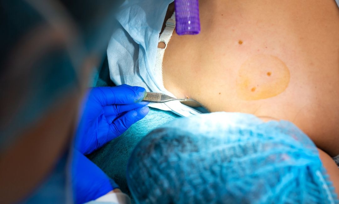
If your dermatologist determines that a mole needs to come off, the good news is that mole removal is a straightforward, in-office procedure. It’s quick, it’s done under local anesthesia, and most people are back to their routine the same day.
Common Removal Methods
| Method | How It Works | Best For | Scarring |
|---|---|---|---|
| Shave excision | Mole is shaved at skin level with a surgical blade | Raised, non-cancerous moles | Minimal, flat scar |
| Surgical excision | Mole and margin of surrounding skin are cut out with a scalpel, then stitched | Suspicious or deeper moles | Small line scar |
| Punch biopsy | A circular tool “punches” out the mole | Small moles requiring full analysis | Small, round scar |
Regardless of the method, the removed tissue is sent to a pathologist for a biopsy to confirm whether the cells are normal or cancerous.
What to Expect After Removal
- First 24 hours: Some tenderness, minor bleeding, and swelling are all normal
- First week: Keep the area clean and dry. Apply ointment as directed. Stitches from excision are typically removed within 7-14 days.
- Weeks 2-4: The wound heals and new skin forms. Avoid picking at any scab that develops.
- Months 1-12: The scar gradually fades. Protect the area from sun damage to prevent darkening.
Reasons for Mole Removal
Moles aren’t always removed because of cancer concerns. Some people choose removal for cosmetic reasons, like a prominent mole on the face. Others remove moles that catch on clothing, irritate during shaving, or simply feel uncomfortable. Freckles and skin tags, while different from moles, are also commonly addressed by dermatologists during these visits.
Pro tip: Always have moles removed by a board-certified dermatologist. If a mole turns out to be cancerous, a dermatologist ensures proper margins are taken during removal, which is critical for preventing the cancer from spreading.
Stay Ahead of Skin Changes With Natura Dermatology
Most moles are completely harmless. But the ones that aren’t can be serious if you ignore them. The key is knowing your skin, checking it regularly, and acting fast when something looks off.
Key takeaways:
- Moles form when melanocytes cluster together instead of spreading evenly across the skin
- Common, atypical, congenital, and blue moles each carry different risk levels
- Use the ABCDE rule monthly to monitor for changes in size, shape, color, and borders
- See a dermatologist immediately if a mole bleeds, itches, or evolves quickly
- Annual full-body skin exams are essential, especially if you have fair skin or a family history of melanoma
- Mole removal is a quick, low-risk procedure that can provide life-saving early detection
Natura Dermatology & Cosmetics was founded on a mission: no one should die from melanoma. Our Fort Lauderdale team performs comprehensive full-body imaging and skin exams designed to catch suspicious moles before they become a threat. If something on your skin has you wondering, don’t wait.
FAQs
What causes moles on the skin?
Moles occur when pigment-producing cells called melanocytes grow in clusters rather than distributing evenly. Genetics and sun exposure are the two biggest drivers. People with fair skin or a family history of moles are at higher risk of developing more.
Do skin moles ever go away?
Yes. Moles typically fade over time, especially after age 50. Some disappear entirely. However, if a mole vanishes suddenly or changes rapidly over a few weeks, have a dermatologist evaluate it. Gradual fading is normal. Rapid changes are not.
Are skin moles good or bad?
Most skin growths classified as moles are completely benign. Having moles is normal, and hair growing from a mole is actually a sign that the skin beneath is healthy. That said, people with many moles or atypical moles do carry a higher risk of melanoma and should monitor them closely.
What do cancerous moles look like?
Cancerous moles are often asymmetrical, have irregular borders, display multiple colors, and measure wider than a pencil eraser. They may also evolve in size or shape over time. Any mole that starts bleeding or becomes painful should be examined immediately.
Should I remove moles?
Not all skin growths need removal. Most moles are harmless and can stay where they are. However, removal is recommended when a mole shows suspicious changes, causes irritation, or when you want it gone for cosmetic reasons. Moles grow back occasionally after removal, so always follow up with your dermatologist.


